How it works
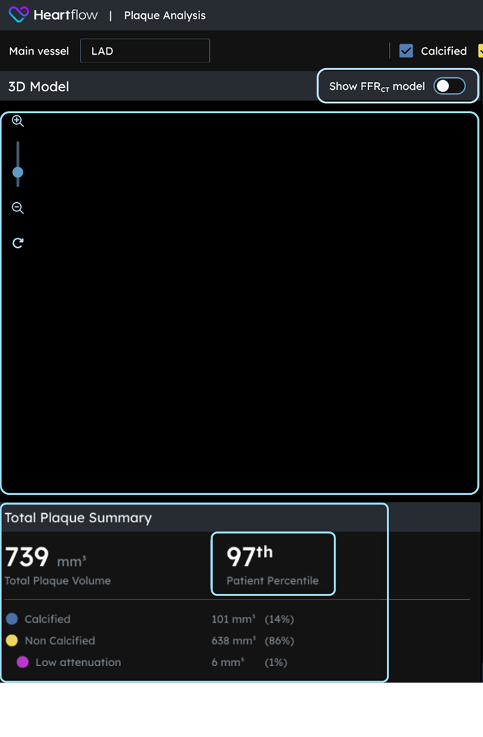
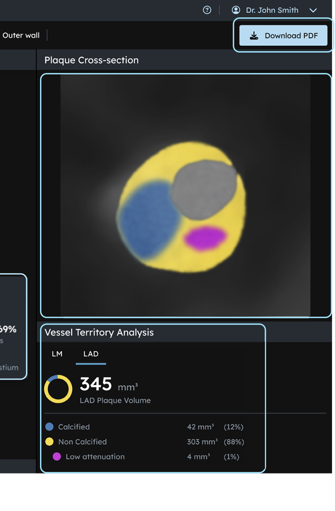
Visualize, Characterize, and Quantify with the Interactive UI.
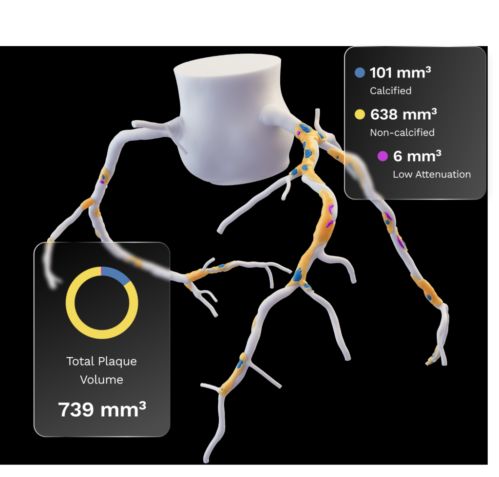
The interactive visualization allows healthcare professionals to explore detailed coronary anatomy and identify potential risk factors with unprecedented precision.
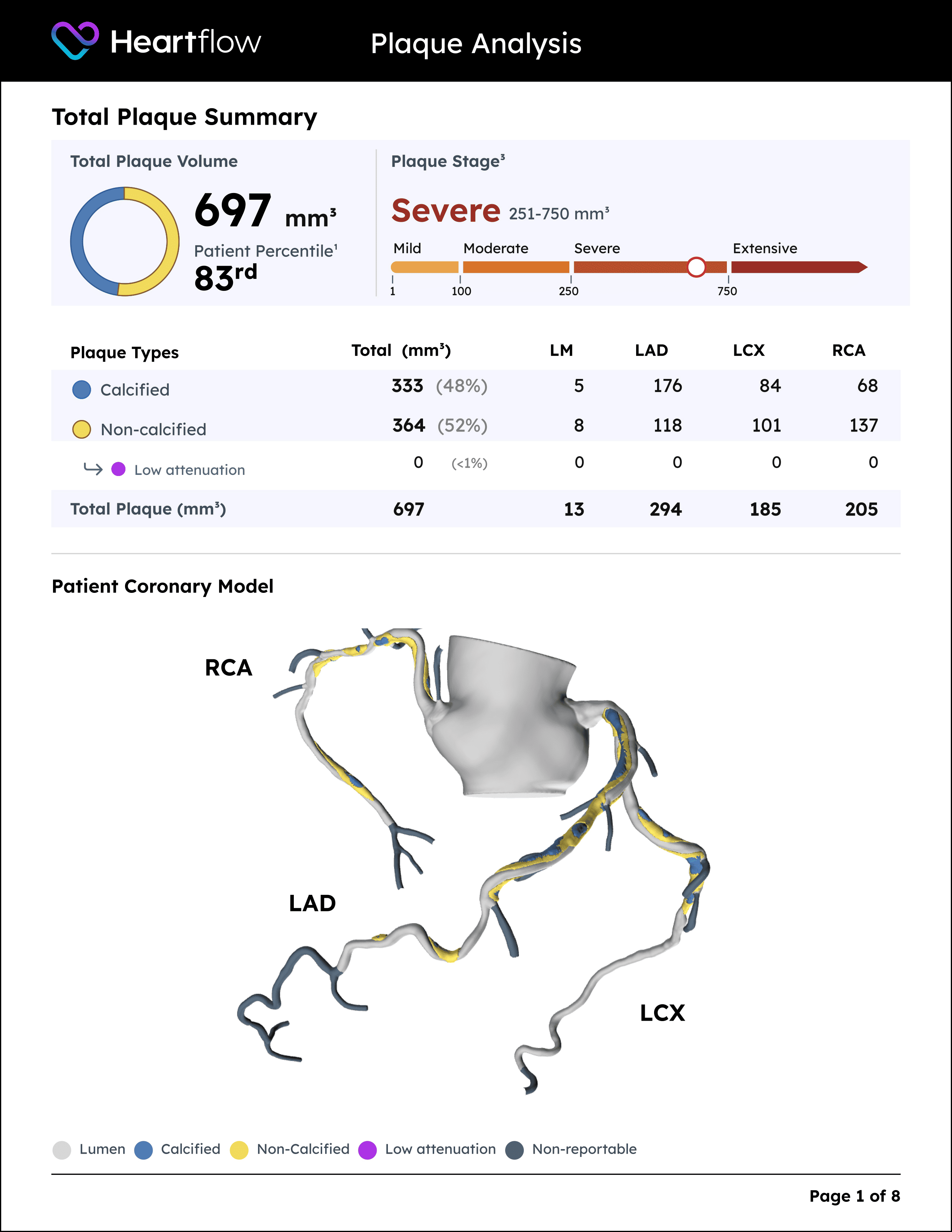
Heartflow Plaque Analysis delivers a comprehensive, AI-driven assessment of coronary artery disease (CAD).

DECIDE is the largest prospective registry on Plaque Analysis in CAD2, demonstrating how Plaque Analysis with Heartflow Plaque Staging empowers physicians with clinical insights that lead to real-world impact.
What if 50% of your CCTA patients could benefit from an adjustment to their treatment plan?
Plaque Analysis identifies and quantifies all plaque types, providing you with personalized data and Plaque Staging insights to determine which 50% of your patients require changes to their treatment plans.

In DECIDE, more than

of patients had a change in medical management after Plaque Analysis.2


agreement with IVUS in precisely quantifying and characterizing coronary plaque.
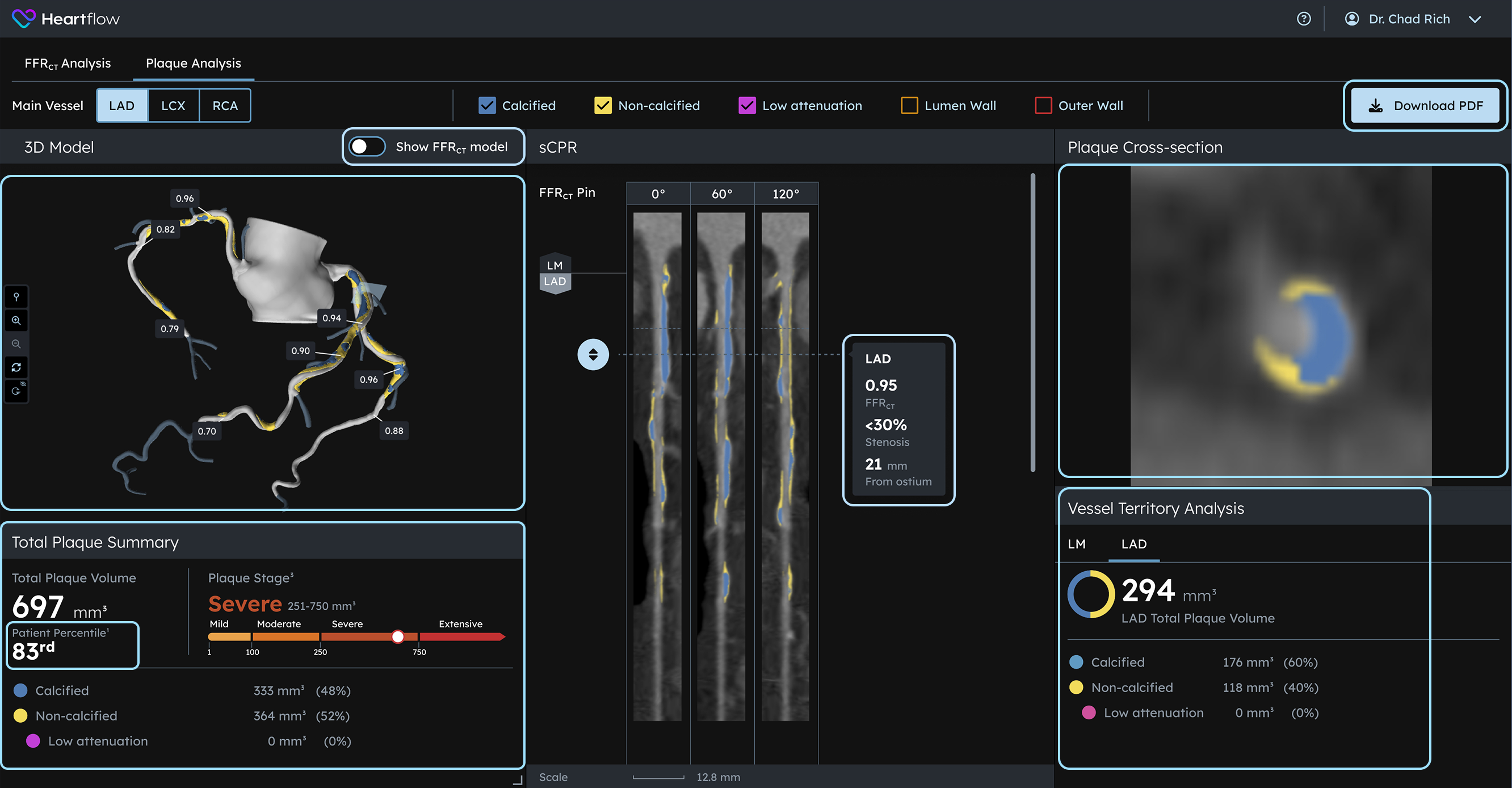
Visualize, Characterize, and Quantify with the Interactive UI.
The interactive visualization allows healthcare professionals to explore detailed coronary anatomy and identify potential risk factors with unprecedented precision.

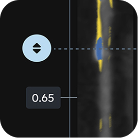
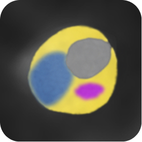
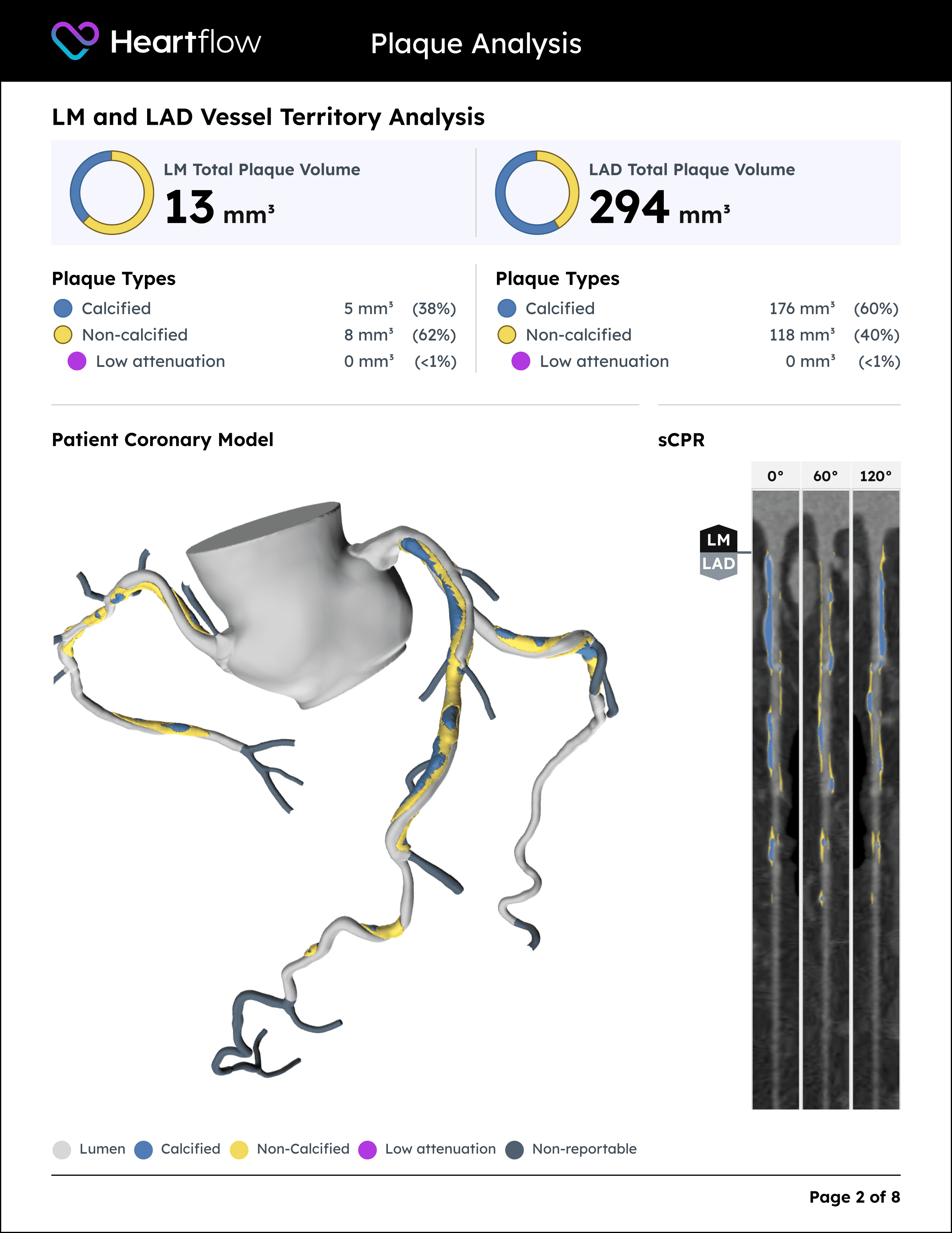
The 3D model, straightened vessel, and cross-sectional views are anatomically co-registered.

If you don't understand the disease, you don't understand the treatment plan. And this kind of addresses both of the paradigms that are very difficult for physicians and patients alike.
”
Discover how you can use Heartflow to advance your approach to coronary artery disease diagnosis, treatment and management.